It was a hot sunny day in southern California when I received a phone call to do a field postmortem. I immediately said yes – I like to do those farm visits because they give me a chance to see the surrounding area and to meet local producers and veterinarians.

The clinician who called me in is a native Californian, a down-to-earth cow veterinarian with a lot of common sense and more than two decades of experience. He left a message on my phone telling me about the case.
At a Holstein calf ranch with increased calf mortality – three calves died overnight. He was on his way to the ranch with some students. I picked up my equipment, one sharp knife, some plastic bags and a bottle of formalin, which is used to preserved samples for microscopic examination.
I then headed down to my car. When I arrived at the calf ranch, the cow vet and three veterinary students, as well as another veterinarian who is interested in animal welfare, were already there.
It was relatively early in the morning, but the three calves were lying in the sun under a cloud of flies. We were told that the calves were 2 weeks old, and they looked small. We placed the first calf on her left side – as I always recommend for consistency – and methodically started the postmortem exam.
The sun was moving up in the sky and it was getting hot, but we managed to finish the first examination without having to rush to beat the heat. The calf had some changes in her gastrointestinal tract and some affected areas in the lung.
By then it was getting uncomfortable under the sun, so we decided to move the remaining carcasses under the shade of a big tree to continue working there. The second and third calves were opened by the students, who were happy for the opportunity to have a hands-on experience.
The second calf had changes similar to the first one. Due to the gastrointestinal pathology, we wanted to rule out the possibility of an enteric pathogen, so samples were taken for submission to the diagnostic laboratory for virology and microbiology testing.
Incidentally, the third calf had an interesting finding. The intestine of this calf did not develop normally; it had a defect called atresia. Atresia means that a segment of the lumen of a tubular organ, like the intestines, was not open during development. In this animal, a section from the colon to the anus was affected.
At times when you do a postmortem exam, you need to be cognizant that some animals may have changes related to the problem you are interested in trying to solve, while some animals will have lesions of no significance to the case.
Atresia ani is a common malformation in newborn calves that may affect as many as 1 percent of newborns but, as in this case, it is not the pathology that led to the death of the affected calf.
 Postmortem examination. Photo courtesy of Ana Alcaraz.
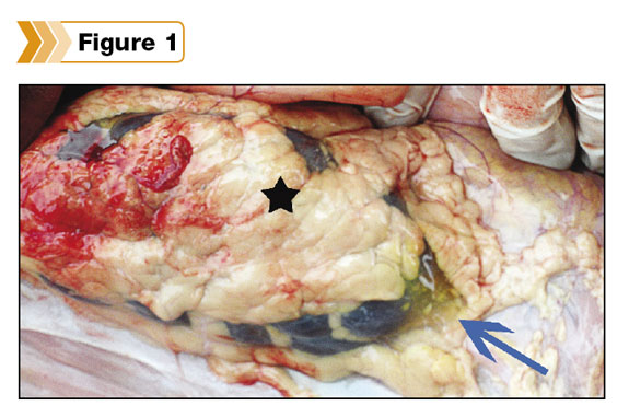
So what was the main cause of death in these calves? We noticed that the three animals showed different degrees of fat around the kidneys and the heart. (See Figure 1.) The presence or absence of fat around certain internal organs is often used to assess the adequacy of the nutrition level of an animal.
The amount of adipose tissue, or fat, found in the postmortem examination should be assessed as adequate or inadequate and determined whether it is excessive or has a different consistency.
In these cases, interestingly, the adipose tissue showed some changes and looked like a clear, gelatinous, soft material, instead of presenting the yellow, firm appearance of normal fat.
The amount of fat was adequate, but its consistency or quality gave us clues that helped us resolve the case. The finding of a gelatinous, soft, clear tissue (See the arrow in Figure 1.) replacing the normal fat stores makes you think of an animal in negative energy balance.
A young animal that is not getting enough energy while it is yet growing very fast starts to consume and use more energy than it ingests and ends up using its internal fat as a source for energy. We recommended the producer test the quality of the milk being fed to these calves.
A field assessment of milk quality was conducted with the help of a Brix refractometer which uncovered a low level of protein in the milk, lower than what calves at this stage in development should be getting.
Based on the results of the postmortem examination, the absence of positive results from the diagnostic laboratory and supported by the low level of protein in the milk being fed to the calves, a diagnosis of what some pathologists jokingly call starvation “virus” was made.
In reality, this is a non-infectious disease, where no actual virus is involved, but it is characterized by young animals growing too fast on a deficient diet which results in atrophied adipose tissue when the animal uses its fat stores as a source of energy.