


A busy postmortem room is a very exciting, entreating environment. It is also an important diagnostic tool for a practitioner and a dairy farmer.

I remember a case coming from a large dairy farm in the Northeast. The calves were kept in hutches, and they were given milk replacer with fluids. The history was brief: The calves were not doing right.
Some calves were regressing from bucket to bottle, and because of that some of them had been force-fed. The farmer and the practitioner sent three calves to the pathology service.
We started working on them early in the afternoon. We opened them with the idea of submitting samples to the diagnostic lab to see what was happening and identify the problem affecting the calves.
When we do a postmortem exam or necropsy, we open three cavities in order to have an overview of what we are getting into. The three cavities are the abdomen, then the thorax and last but not least, the heart, which is called the pericardial cavity.
It is at this moment, when we are observing the whole inside of the animal, that we decide if we need to call for the support of the last member of a typical diagnostic team: the laboratory.
When I send tissues to the diagnostic lab, I need to know how they are going to help me explain the results. In addition, I need to understand why I am sending the samples. Maybe we want to know the cause of the changes found during the postmortem.
In cases when we are unable to observe any macroscopic changes during the postmortem, we have to make use of all of our experience – because the animal could have died so fast that there is nothing to see with the naked eye.
In an exam, we consider the species, the visible signs of health (or lack thereof), the manner of death and the history associated with the case in order to gain an idea of what we are looking for. In these situations, like in many others, the diagnostic lab will help us to identify the culprit.
In the case I began describing earlier, we found that two of the three animals that we received had changes in the abomasum and omasum, and the third one had no changes.
Despite knowing these calves’ history of not doing well, having some diarrhea and not wanting to eat, the third calf’s lack of internal changes was puzzling. Did all three die of an underlying disease? Did two succumb to something different than the cause of the third’s demise?
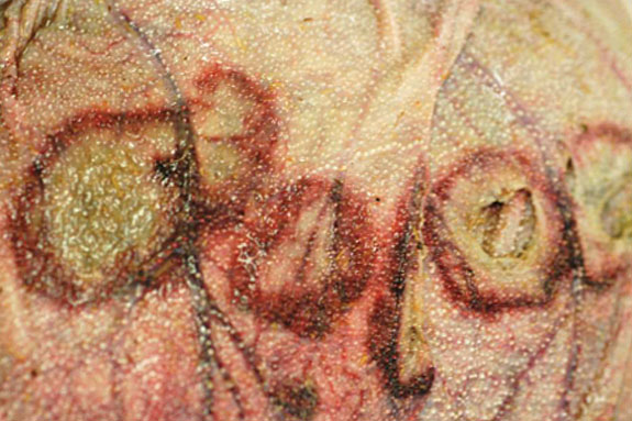 This photo shows multiple target lesions in the omasum. You can see the target pattern of the colors.
The findings in the abomasum and omasum in the first two calves were multifocal, well-demarcated areas with a peripheral red ring and a central pale area.
Some pathologists describe these changes as target lesions, like a bull’s eye, because of the shape and particular colors.
These changes are usually associated with a fungal problem, or mycotic diseases.
Fungal organisms are opportunistic agents, which means these organisms are everywhere in the environment, just waiting for the right opportunity to take over a weak animal.
When the animals are debilitated or weakened by a primary viral or bacterial infection, the fungal organisms find a chance to proliferate and take over.
Even though the exam indicated an opportunistic fungi, it is important to identify the underlying bacterial or viral pathogen that weakened the animal to start with, as those pathogens also need to be brought back under control.
We took samples for the diagnostic lab because, after seeing the lesions, our first suspect was the possibility of a secondary, fungal, opportunistic disease. In order to identify the primary pathogen, during the necropsy we took tissue samples to look for a virus or a bacteria or maybe a parasite.
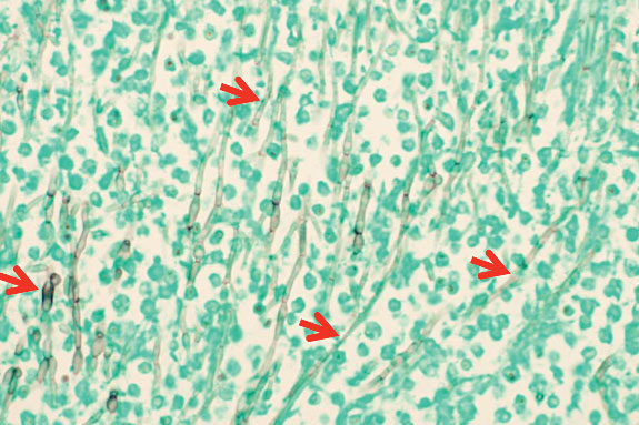 A microscopic picture of the abomasum with numerous perpendicular rows of fungal organisms, also called hyphae. (See red arrows.) The green background is the tissue.
A week after the postmortem examination, with the help of the microscope, we found inflammation of the intestine and the presence of bacteria and some parasites.
The omasum was completely covered by fungal organisms and when special stains were used, the fungi were stained in black.
Since the fungi are opportunistic agents, we still waited for the lab to identify the primary cause which lead to the animal’s weakening and permitted the proliferation of the fungi.
It was not surprising that the diagnostic lab found bacterial disease (E. coli), viruses (coronavirus) and parasites (cryptosporidium) in all three. The debilitating state of these animals made the right environment for the fungal organism to grow, proliferate and take over the omasum in two of the animals.
The third succumbed to those same pathogens alone. It is important to remember that the same disease process may present itself differently in different animals and under different circumstances.


