Editor’s note: This is the first in a two-part series. To read part two click here.
People rarely think the way they manage a difficult birthing situation could determine the potential for injury to the cow and calf resulting in temporary or even permanent lameness. In the following article, we offer some important considerations for those who offer assistance to cows requiring assistance at calving. The delivery techniques applied have important implications for both the cow and calf.
Dystocia-related injuries to cows and calves
Dystocia is defined as difficult birth, and the causes are many, but most calving problems are related to abnormalities in the fetuses’ presentation, posture, position and its size relative to the cow’s pelvis.
Fetal presentation
The “presentation of a fetus,” in obstetrical terms, is determined by its spinal axis relative to that of the dam. It may be longitudinal (i.e., up and down) or in a transverse (sideways) plane in relation to how the fetus enters the birth canal.
Most fetuses enter the birth canal head-first with the front legs extended forward as if diving out of the birth canal. This is referred to as anterior presentation.
Sometimes, fetuses will be presented to the birth canal with their back feet and legs coming first. This is referred to as a posterior presentation. On fortunately rare occasions, a fetus may enter the birth canal with the spine or all four feet presented first.
Just to complicate things still further, the fetus may be in longitudinal or transverse orientation. In either case, these abnormal presentations represent some of the most complicated obstetrical challenges and are best left to the experience of a veterinarian.
Now consider the complications to those described above when the limbs or feet of more than one fetus are simultaneously presented into the birth canal (i.e., such as occurs with the delivery of twins). Before any attempt is made to extract a fetus in this situation, one must first determine if the limbs belong to one or more than one fetus.
If we assume this predicament is a consequence of twins, and one of the fetuses is in an anterior presentation (i.e., front legs and head coming first as if diving out of the birth canal), and the other in posterior presentation (i.e., rear legs and buttocks are coming first), the first thing to do is to distinguish front legs from rear legs. Simple, right – or is it?
For anyone who has been in this situation, they’ll know that when working blindly within the uterus of the cow, rear feet (to the level of the fetlock joint) of the fetus are indistinguishable from front feet. The same is true for the hock and elbow joints.
Sorting this out requires that the obstetrician count the number of joints that flex backward from the foot to the elbow or hock joint which both flex forward.
For example, starting with the foot and working upward on a front limb, the operator encounters two joints: the fetlock and the carpus or knee, both of which flex backward, before finally reaching the elbow joint, which flexes forward.
It is important to never apply traction until it is clear that both feet belong to the same fetus and that both leg holds are applied to either front feet or back feet. If one of two feet is a rear foot and the other a front foot, it may be twins or worse yet a longitudinal or transverse presentation. Experience is the secret to developing confidence with these techniques.
Fetal position
The “position of a fetus” refers to its spinal orientation relative to the dam’s. In other words, the position is incorrect when the fetus is upside-down (belly of the fetus is facing upwards toward the cow’s spine) when entering the birth canal.
When a fetus is coming backwards (i.e., posterior presentation) and the feet become visible beyond the vulva, the fetus will appear to be upside-down.
On the other hand, the feet of a fetus in anterior presentation that is upside-down will give the appearance that the fetus is coming backwards. Since dystocia is more common in fetuses coming backwards (i.e., posterior presentation), early intervention is recommended.
Rarely, a fetus may be upside-down (belly of the fetus on the sky side) and backwards, which makes it appear to be coming normally in anterior presentation.
It is easy to become fooled by these observations, so it is important to understand these relationships when managing calving situations. Possibly one of the best tools for managing calving situations is a wristwatch.
When the fetus (based upon the appearance of feet at the vulva) appears to be in anterior presentation, continue to monitor the cow closely. If feet are visible at or slightly beyond the vulva, the fetus should be delivered within an hour. If it is not, an examination is in order.
Fetal posture
Finally, the “posture of a fetus” refers to the extremities of the fetus, which may be flexed, extended or otherwise retained above, below or to one side or another. For example, when one leg is retained or the head is turned to one side, the posture of the fetus is incorrect.
In dystocia situations, the objective is correction of the presentation, position and posture of the fetus so that it may be delivered naturally. When the fetus is too large compared to the dam (particularly the dam’s pelvis), delivery may be prolonged or impossible without assistance.
In Part II, we continue this discussion with a look at how to determine if a calf is too large to be delivered by forced extraction (pulling). We’ll also take a closer look at common lameness disorders in cows and calves occurring from dystocia. ![]()
PHOTOS
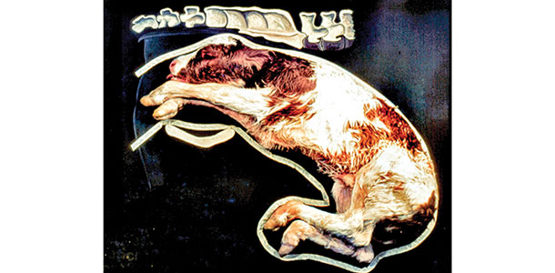
PHOTO 1: Posture is the relationship of the calves’ extremities with respect to the body. In this case the front leg is held back.
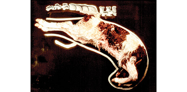
PHOTO 2: Anterior presentation.
PHOTO 3: Posterior presentation. Photos courtesy of the Drost Project and drostproject.org

- Jan K. Shearer
- Professor and Extension Veterinarian College of Veterinary Medicine
- Iowa State University