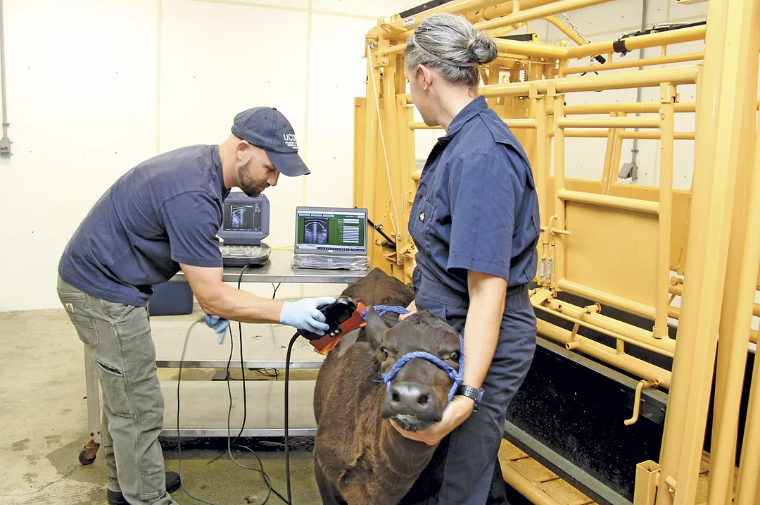
Mostly known for its use in reproductive work – confirming pregnancy, looking at fetal gender, fetal age and ovaries – ultrasound technology has many uses in the beef industry.

With a change of a probe, ultrasounds have become a standardized measuring tool for assessing an animal’s carcass traits, conducting research and medical imaging. The portable equipment has the same high-quality capabilities as human products but is also durable and rugged enough to be used outdoors, in dusty barns or corrals.
Carcass evaluation
Made up of researchers and industry reps, the Ultrasound Guidelines Council (governing body for ultrasound work globally) enables breed associations to standardize ultrasound data to check backfat, ribeye area and intramuscular fat.
Two main ultrasound interpretation labs are used – Centralized Ultrasound Processing Lab (CUP) in Ames, Iowa, and UltraInsights Processing Lab in Pierce, Colorado. Kevin Gould, an extension beef educator at Michigan State University, says, “We’ve [used ultrasound] mainly with seedstock the past 30 years, trying to increase predictability before we utilize animals for breeding. We determine actual breeding values for those three carcass traits.”
Technology today is improving for measuring high marbling scores. Gould says, “The new equipment gives a better range of ultrasound interpretation for marbling scores across a contemporary group and expands the range of data so you can identify outlier animals easier."
Outliers can be good or bad. It depends on the trait and your goals and the environment you raise cattle in. “It allows us to focus on what the value is in the marketplace for the cattle we raise, to try to get paid for that value, and gives a better way of interpreting that data,” Gould says.
“Genomics came on the scene in the past 10 years. Ultrasound and genomics are complementary tools. Genomics measures the animal’s genes, and we try to interpret how those genes will affect certain traits we’re trying to select for, or against," Gould explains. "With genomics, we can look at parentage, lethal defects and other things that ultrasound can’t. What ultrasound can do is measure the animal’s actual data for that trait, in the environment it’s raised in, and compare it within the contemporary group it’s being raised with."
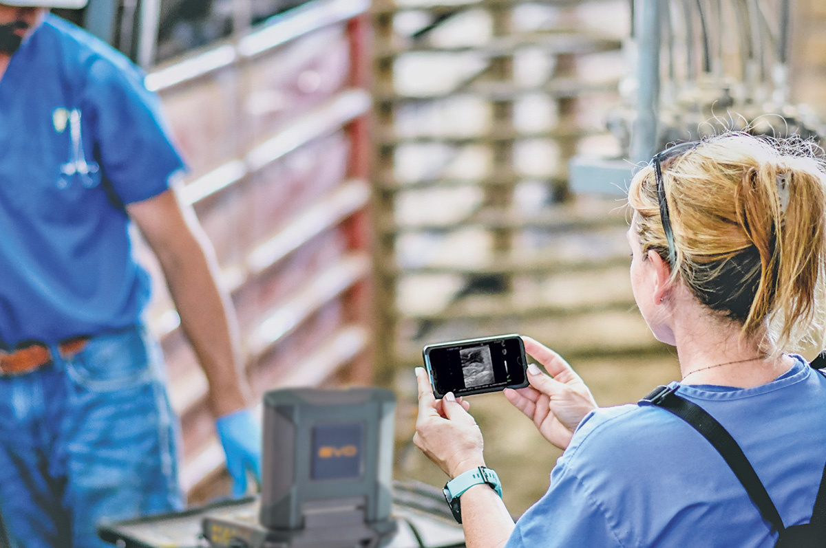 A bovine ultrasound exam can be streamed to the phone of anyone nearby; they can
be at a safe distance from the actual exam being done. Photo courtesy of Erika Wierman.
A bovine ultrasound exam can be streamed to the phone of anyone nearby; they can
be at a safe distance from the actual exam being done. Photo courtesy of Erika Wierman. Larger contemporary groups generally show a larger range in each trait and help in indexing animals within the group. “We find the outliers; the complementary benefits of both technologies are very good. The more accuracy we have for both, the more information we have and the better decisions we can make,” Gould says.
Ultrasound data from cattle scanned at the farm goes to the lab, where it is interpreted and all data are ranked for each trait for each animal, under its registration number. That information is sent to the breed association, to influence that animal’s expected progeny differences (EPDs). “This increases accuracy of EPDs for each trait we scan for [backfat, ribeye area and marbling score],” he explains.
Traits can be measured in feedlot animals (to predict harvest timing) and with accuracies above 80% in live animals, representing what the actual carcass data on the rail would be.
If breeding heifers are scanned, we can find out if they really belong in the herd. “We need to know ahead [of time], rather than find out later that they are not what we wanted,” Gould says.
Gould also scans cattle for youth programs and research. He recently scanned 50 market steers from the county fair for a carcass contest and ranked them on the USDA grid, so the highest-value carcass can win. This gives kids an opportunity to learn about carcass merit and an aspect of the industry they rarely get to see.
Determining the best feeding program
Joe Emenheiser, a livestock extension educator at the University of Connecticut, is involved with research utilizing ultrasound to evaluate beef-on-dairy cross calves. This research is a collaboration between faculty at UConn and Penn State.
“Our team is looking at effects and economics of different management practices with these calves, since many dairy producers are now breeding some of their cows to beef sires. We’re evaluating two different calf milk replacer treatments and two grower rations to evaluate how calf performance changes – depending on level of nutrition and investment. Here in the Northeast, many dairy-cross calves are fed dairy cow refusals [waste from the TMR (total mixed ration) scraped from the bunk after dairy cows consumed most of it] or fed like dairy heifers. Neither method is very appropriate for a beef calf,” Emenheiser says.
Finding the best way to feed and raise these calves to optimize eventual carcass quality is the premise for the project, with the aim of improving growth and carcass composition – and final meat quality.
“We’ll be evaluating different carcass measurements, tracking this through their lifetime to develop growth curves and estimates of when the most appropriate time to harvest them might be and how that changes with different treatments [nutrition]. We ultrasound all UConn crossbred calves beginning at [1] month of age, and every month thereafter until harvest. This helps us evaluate the effects of different nutrition protocols, along with data on calf performance, carcass value and cost of production,” he explains.
As opposed to a genetic evaluation, which is done just one time on a yearling bull or heifer, Emenheiser says they are trying to build descriptive growth curves for the different carcass composition traits – loin muscle size, backfat thickness and percentage of intramuscular fat or marbling.
“We want to see if there are developmental differences between calves that are managed differently, and whether there is a more optimal time to harvest them – relative to carcass yield and quality criteria – with these different approaches,” he says. It might be possible to tweak the management and nutrition to achieve target goals more optimally.
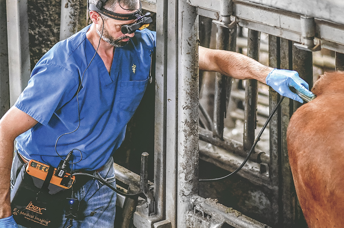 The operator doing the exam
with goggles can see the exam without having to look at a screen, and a video
can also be made from the ultrasound. Photo provided by Erika Wierman.
The operator doing the exam
with goggles can see the exam without having to look at a screen, and a video
can also be made from the ultrasound. Photo provided by Erika Wierman.
Ultrasound for diagnostics
Erika Wierman, DVM, staff veterinarian for E.I. Medical Imaging, says that in addition to the new portable laptop ultrasound units that can be set up chuteside and waist-worn, goggle-based units that provide a self-contained, mobile setup (with options for streaming the image from the ultrasound to mobile devices or a larger monitor, TV or projector) have also improved veterinarians’ ability to diagnose many problems – including in the lungs.
“We are often screening calves and looking at lungs for signs of early or subclinical respiratory disease. If we can identify and treat those calves earlier, they recover quicker and make better gains, with fewer issues down the road,” Wierman says.
Ultrasound is now a widely used tool for looking at the outer surface of the lungs (pleura), especially in calves susceptible to pneumonia in feedlots. Since ultrasound waves don’t penetrate through air, you can’t get a full picture of the lungs like you would with radiographs. “But we can look at the quality – normal versus abnormal – of the surface of the tissue-air interface. The lung surface underneath the ribs should be smooth and free of any abnormalities,” she says.
“With ultrasound, we can identify irregularities on that surface, such as abscesses or areas where tissue is consolidated and unable to fill with air. We can detect places where there are indications of disease,” says Wierman. This type of examination can also help people determine which calves are sick enough to not keep putting more money into treatment and feed.
Veterinarians also use portable ultrasound to evaluate teats for mastitis; evaluate musculoskeletal injury, abscesses and other superficial anomalies; and for transabdominal applications such as ultrasound-guided biopsies of the liver.
“There are several new studies starting, and an old study being revised, looking at curd formation in calves’ stomachs. This is mostly in dairy calves, but it illustrates the fact that ultrasound can be used to look at the forestomach,” Wierman says.
Ultrasound can also be a diagnostic tool for hardware disease. “You could potentially look in the front part of the abdomen for abnormalities like that.” The veterinarian might suspect a problem with one of the stomachs, like an impacted abomasum, and ultrasound might be helpful.
There are many uses for ultrasound in internal medicine, and new uses for this technology may continue to come along.